Peyronie's Disease
Jump to navigation
Jump to search
Includes 2015 AUA and 2018 CUA Peyronie's Disease Guideline Notes
Definition[edit | edit source]
- Criteria (3):
- Acquired penile abnormality
- Characterized by fibrosis of the tunica albuginea
- Which MAY be accompanied by (4):
- Pain
- Deformity, including curvature, indentation, hinge effect, and shortening
- Erectile dysfunction
- Distress
Epidemiology[edit | edit source]
- Prevalence: 9% of men
- Peak age of onset is early 50s
- Affects all races
Pathophysiology[edit | edit source]
- Peyronie's disease (PD) is an inflammatory disorder of the tunica albuginea
- Exact cause not known.
- Some injury to the erect or flaccid penis is necessary to trigger the cascade of events that leads to PD in the susceptible individual.
- Microvascular trauma to the penile shaft associated with penile buckling in the erect or semi-erect state secondary to sexual activity is thought to be the most common inciting event
- Most males cannot recall a specific event
- Not all trauma is associated with PD
- Microvascular trauma to the penile shaft associated with penile buckling in the erect or semi-erect state secondary to sexual activity is thought to be the most common inciting event
- In PD, the scar does not undergo a normal remodeling phase in the wound healing process and therefore the scar and deformity persists
- Normal wound healing involves 3 phases:
- An acute phase
- A proliferative phase
- A remodeling phase
- Normal wound healing involves 3 phases:
- Some injury to the erect or flaccid penis is necessary to trigger the cascade of events that leads to PD in the susceptible individual.
- Exact cause not known.
Risk factors[edit | edit source]
- IT DRAG A Crooked Wand (9):
- Infection
- Trauma
- Diabetes
- Radical prostatectomy (prevalence 11-16%)
- Increasing Age
- Genetic predisposition
- Autoimmune factors
- Collagen disorders (Dupuytren contracture, fibrosis of plantar fascia (Ledderhose’s disease), tympanosclerosis)
- Aberration of localized Wound healing
- Others: hypertension, hyperlipidemia, smoking, heart disease
- Hypogonadism is associated with more severe Peyronie's disease.
- Supplemental testosterone improved the efficacy of intralesional verapamil in patients with hypogonadism
- PDE5 inhibitors are not associated with Peyronie's disease
Cellular physiology[edit | edit source]
- Oxidative stress has a well-documented role in tissue fibrosis and has been studied in the pathogenesis of PD
- Nitric oxide has anti-fibrotic effects
- Myofibroblast activation is a key event in the development of fibrosis
- TGF-β1 has been shown to be significantly associated with PD. TGF-β is a strong activator of myofibroblasts and is known to be a potent fibrotic growth factor by stimulating the deposition of ECM.
- A variety of alterations may be present in a given patient so no treatment works uniformly for all patients
Penile Anatomy and Peyronie's Disease[edit | edit source]
- See Penile Anatomy Chapter Notes
- Tunica albuginea
- Tough connective tissue layer composed primarily of type I collagen
- A bilayered structure (outer longitudinal, inner circular) with multiple sublayers.
- Outer longitudinal layer
- Thinnest at the 3 and 9 o'clock positions of the corpora
- Completely absent on the ventral groove between the 5 and 7 o'clock positions.
- The most vulnerable area in the tunica albuginea
- Most prostheses tend to extrude here
- The lack of tunica albuginea on the ventral groove may contribute to greater ease of dorsal buckling and explain why most PD patients exhibit dorsal curvature
- 60-70% of plaques are located on the dorsal aspect of the penis and are usually associated with the septum
- The most vulnerable area in the tunica albuginea
- Outer longitudinal layer
- Covers the corpora cavernosa and *spongiosum
- *The corpus spongiosum lacks an outer longitudinal layer or intracorporeal struts, ensuring a low-pressure structure during erections
- Penile deformity is caused by asymmetrical expansion of the corpora.
- When expansion is limited at one point along the circumference of the corpora by the inelastic scar of the Peyronie plaque, deviation to affected side occurs
- Note that in penile fracture, the penis deviates away from the affected side (side of injury)
- A circumferential plaque may lead to an hourglass deformity
- When expansion is limited at one point along the circumference of the corpora by the inelastic scar of the Peyronie plaque, deviation to affected side occurs
Natural history[edit | edit source]
- Characterized by symptoms with a variable course, some of which may improve or resolve without treatment in some patients.
- Plaque-related pain improves and/or resolves in the majority of patients with time even in the absence of treatment. Conversely, spontaneous resolution or significant improvement of penile deformity is rare
- The natural history of Peyronie disease is complete pain resolution in ≈90% of men
- Penile curvature improves in only 12-13% of men, worsens in ≈45%, and remains stable in ≈42%.
- Active vs. Stable Disease
- Active (acute) disease:
- Characterized by dynamic and changing symptoms
- Defining symptom: penile and/or glanular pain or discomfort with or without erection
- Can last up to 18 months[1]
- Stable (chronic) disease:
- Symptoms have been clinically quiescent or unchanged for ≥3 months
- Pain with or without erection may be present but is less common
- Additional symptoms include difficulty in maintaining erectile function and inability to sustain intercourse
- Active (acute) disease:
- The presence of penile pain, especially with erection, increases the likelihood of a penile septal scar, an atypical form of Peyronie disease, which may be characterized by symptoms including penile shortening, pain, and ED, even in the absence of deformity or palpable disease
Differential diagnosis (4)[edit | edit source]
- Congenital penile curvature
- Thrombosed or torn dorsal penile vein
- Penile fracture
- Penile cancer (rarely)
Diagnosis and Evaluation[edit | edit source]
UrologySchool.com Summary[edit | edit source]
- AUA:
- Mandatory (1): history and physical exam
- Recommended (1): detailed examination of erection (photograph or in-office ICI testing) prior to invasive intervention
- CUA:
- Mandatory (1): history and physical exam
- Recommended (1): detailed examination of erection (photograph or in-office ICI testing) prior to invasive intervention
History and Physical exam[edit | edit source]
History[edit | edit source]
- Signs and Symptoms
- Onset, duration, acquired vs. lifelong (to differentiate PD vs. congenial penile curvature), changes over time (deformity and erectile function)
- Penile characteristics: extent of penile deformity, direction of curvature, palpable plaque(s), presence of hourglass deformity, shortening, and presence of hinging, penile pain with and without erection. Penile sensation, ejaculatory function, and length/girth concerns should be documented.
- Pain, when present in the acute phase, can occur in the flaccid penis with palpation of the plaque, with erection, or during intercourse
- Once the disease process is stable, most pain will resolve, but in some men the pain persists with a pulling sensation on the plaque when a strong erection occurs (torque pain)
- Risk factors
- History of traumatic event
- Medical history inclusive of family presence of Peyronie disease, Dupuytren’s contracture, and other related conditions that may impact erectile and sexual function.
- Sexual function: erectile rigidity, interference with intercourse, ability to penetrate, ability to complete intercourse, and partner complaints and support should be documented. The use of the International Index of Erectile Function-5 (IIEF-5) or the Disease Questionnaire Peyronie’s (PDQ), which is a newer, 15-item, validated instrument specific to Peyronie disease may be of use.
- Psychologic impact: Many men with PD experience emotional distress, depressive symptoms, and relationship difficulties. PD can have a profound negative impact on men’s QoL.
- Based on bother/psychologic impact, consideration may be made for referral to a mental health professional with expertise in sexuality.
- Prior Peyronie disease and ED treatments should be documented.
- AUA Guidelines: “The recent onset of penile curvature and varying degrees of penile pain, without a palpable penile abnormality, in the non-erect state, may be considered diagnostic”
- A diagnosis of ED or failed first- and second-line ED treatments warrants ruling out Peyronie disease
Physical exam[edit | edit source]

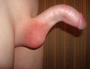
Patient with ventral penile curvature. Source: Wikipedia
- Penis
- Should be examined on stretch
- Assess for palpable abnormalities
- Plaque(s) can be palpated or documented on ultrasound.
- Identify the location, size, number and tenderness of the plaque
- Most common location of plaque is on the dorsal mid-shaft
- Measurement of size of the plaque with any modality, including imaging, has been found to be inaccurate
- Prior to any intervention, measurement of stretched penile length, from the penopubic skin junction to the coronal sulcus or the tip is recommended to establish baseline penile length as penile length loss is a primary concern and contributor to distress for Peyronie disease patients
- Digital home photographic documentation may aid in objectively determining treatment effects, especially when non-surgical options are used
Laboratory[edit | edit source]
- Routine laboratory testing is not recommended
- Targeted bloodwork may be obtained in response to specific findings on history or physical examination (for example, signs/symptoms of hypogonadism)
Imaging[edit | edit source]
- Options
- Ultrasound
- Benefits (4):
- Identification and measurement of calcification during initial surveillance in the flaccid state
- Calcification may occur early after the onset of the scarring process, not necessarily only in chronic/mature disease.
- Men with extensive calcification are less likely to benefit from non-surgical treatment
- Identification of corporeal fibrosis
- Assessment of penile vascular flow parameters after intracavernosal injection of vasoactive agent
- Observation of the erectile response to the vasoactive injection compared to the patient’s sexually induced erection; best opportunity to objectively assess deformity (curvature, girth irregularities, hinge effect)
- Identification and measurement of calcification during initial surveillance in the flaccid state
- CUA: colour duplex ultrasonography may be offered, but may not be readily available; combination of ultrasound with intracavernosal injection may also identify arterial insufficiency or veno-occlusive dysfunction, influencing choice of Peyronie disease management
- Insert figure
- Benefits (4):
- MRI
- Excellent imaging modality for penile pathology, including PD
- Routine use in clinical practice is not supported
- CT and plain radiography do not have a role
- Ultrasound
Other[edit | edit source]
- Prior to any invasive intervention (e.g., intralesional treatments or surgery) obtain accurate assessment of erection including (4):
- Erect penile length
- Degree of curvature
- Presence of hourglass deformities
- Rigidity of erection
- Accurate assessment of erection can be obtained with
- Digital photographs
- Examination after intracavernosal injection (ICI) of vasoactive agents, with or without duplex Doppler ultrasound (gold standard)
- The ICI test enables assessment of penile deformity, plaque(s), and pain in the erect state.
- When the ICI test is combined with duplex ultrasound, additional measurements of plaque size and/or density can be made, calcified and non-calcified plaques can be differentiated, and information on the vascular integrity of the penis can be obtained.
- AUA prefers in-office intracavernosal injection (ICI) test with or without duplex Doppler ultrasound but describes that home photography of the erect penis with the use of a protractor during an erection in the office may be sufficient to document deformity from some cases.
- CUA describes both as options but describes the use of ICI as the most reliable method and that penile injection remains the gold standard, especially in patients reporting complex deformity (hourglass or bidirectional curvature) or ED
Management[edit | edit source]
- 2015 AUA and 2018 CUA Guideline Notes are included here
- Only clinicians with expertise in Peyronie’s disease should treat affected patients
- Discuss with the patient a care plan, which is consistent with patient symptom status, current health, and treatment goals.
Options[edit | edit source]
- Observation
- Intervention
- Active phase (1):
- NSAIDs for pain
- Stable phase
- Non-surgical (2)
- Penile traction
- Intralesional injection
- Surgical (3)
- Plication
- Plaque excision +/- grafting
- Penile prosthesis
- Non-surgical (2)
- Active phase (1):
Observation[edit | edit source]
- Patients without pain or difficulty in accomplishing penetrative sex may only require reassurance
Intervention[edit | edit source]
Non-surgical[edit | edit source]
Options (4):[edit | edit source]
- Penile traction
- Oral medication
- Intralesional injection
- ESWT
UrologySchool.com Summary of non-surgical interventions for Peyronie's Disease[edit | edit source]
- Treatment depends on whether the patient’s symptoms are active (dynamic) vs. stable
- AUA:
- Recommended:
- Active phase (1):
- NSAIDs for pain
- Stable phase (2):
- Penile traction
- Intralesional (collagenase, verapamil, interferon alfa-2b) injection with modelling
- Active phase (1):
- Conditional (1):
- Extracorporeal shockwave therapy for pain
- Recommendation is Conditional because the broader PD literature indicates that pain is the PD symptom that is most likely to resolve over time without intervention, the patient burden involved in obtaining ESWT treatment to treat pain may be substantial, and other treatments may be equally effective at alleviating pain. Further, ESWT is associated with frequent adverse events
- Extracorporeal shockwave therapy for pain
- Recommended:
- CUA (mostly similar to AUA, except CUA lists some oral medications that may be considered while these are not supported by AUA):
- Recommended:
- Active phase: NSAIDs for pain
- Stable phase: penile traction, intralesional treatment with modelling (first-line: collagenase; second-line: verapamil or interferon where cost or concern related to adverse events limits use of collagenase; AUA does not use similar hierarchy of preferred treatments)
- May be considered:
- Oral medication (alone or as a part of multimodal care) (different than AUA):
- Can Consider Peyronie's Pills (4)
- Colchicine
- Co-enzyme Q10
- Potassium paraaminobenzoate
- Pentoxifylline
- Extracorporeal shockwave therapy for pain
- Oral medication (alone or as a part of multimodal care) (different than AUA):
- Recommended:
Penile traction[edit | edit source]
- First-line non-invasive, non-surgical treatment modality
- No RCT to date
- Associated with (4):
- Reduced curvature
- Increased stretched penile length
- Increased erect girth
- Correction of hinge effect
- Benefit maintained at 6 months of completion of therapy
- Critical that patient wears device for ≥3 hours per day to get satisfactory results
- Insert figure
Oral medication[edit | edit source]
- Oral non-steroidal anti-inflammatory medications can be used for pain in the active phase
- Tried but not useful/not much evidence: colchicine, co-enzyme Q10, potassium aminobenzoate (Potaba, member of the vitamin B complex), pentoxifylline (increases nitric oxide; RCT trial retracted 6 years later due to inappropriate statistical analysis), carnitine, omege-3 fatty acids, procarbazine, tamoxifen, vitamin E, vitamin E with L-carnitine
- Low-dose daily tadalafil is a safe and effective treatment option in septal scar remodeling, but limited evidence
Intralesional injection[edit | edit source]
- Clostridial collagenase (Xiaflex) with penile modeling
- Produced by the bacterium C. histolyticum
- Selectively degrades collagen types I and III in connective tissues
- Associated with:
- Decrease in deviation angle
- Reduction in plaque size
- Does NOT treat pain or ED
- Appropriate in patients with (2)
- Curvature >30° and <90°
- Intact erectile function (with or without the use of medications)
- IMPRESS I & 2
- 832 PD patients with (4):
- Stable disease
- Use in active phase is considered off-label
- Curvature > 30° and < 90°
- Single dorsal or lateral plaque
- No isolated hourglass deformity or calcified plaque
- Normal erectile function
- Stable disease
- Randomized to intralesional collagenase vs. placebo
- Results:
- In both trials, men in the CCh group were shown to exhibit a 17° improvement in penile curvature compared to 9° in the modelling only, placebo group, modest absolute difference 8°
- 832 PD patients with (4):
- To-date, clinical trials have not evaluated the use of collagenase in patients with (5):
- Hourglass deformity
- Ventral curvature
- Calcified plaque
- Plaque located proximal to the base of the penis
- Curvature < 30° and > 90°
- Adverse events (5):
- Corporal rupture
- Extremely rare; will often necessitate surgical repair
- Penile bruising
- Penile pain
- Penile swelling
- Hematoma
- Corporal rupture
- Should be combined with penile modeling by the physician and patient
- Discontinued in Europe and Canada in 2020 due to cost and poor market uptake§
- Calcium channel blocker
- Verapamil
- Affects fibroblast function on several levels, including cell proliferation, ECM protein synthesis and secretion, and collagen degradation
- Evidence for the use of intralesional verapamil is weak
- Poor candidates include those with:
- Extensive calcification
- Curvature >90°
- Ventral curvature (difficult to adequately infiltrate the plaque)
- Predictors of success include:
- Younger age (age < 40)
- Curvature >30°
- Adverse Events (4):
- Penile hematoma
- Pain
- Dizziness
- Nausea
- Nicardipine
- Demonstrated significant reduction in pain, improvement in IIEF-5 score, and reduction of plaque size when compared to placebo
- Verapamil
- Interferon alfa-2b
- Efficacy
- Used to reduce curvature, plaque size, pain, and improve some vascular outcomes
- Modest improvement compared to placebo
- Indications
- Curvature > 30° and without calcified plaque
- Adverse Events
- Sinusitis
- Flu-like symptoms of arthralgia
- Fevers
- Chills
- Local effects including penile hematoma, swelling, and pain
- Significant cost and side effects
- Rarely used in Canada
- Efficacy
- Corticosteroids
- Not recommended for intralesional treatment of Peyronie disease
- Hyaluronic acid and botulinum toxin A (onabotulinum toxin A)
- Too early to make any recommendations on the use of these medications until more safety and efficacy data are available
- Platelet-derived growth factors (platelet-rich plasma)/Priapius ShotTM protocols and stem cell therapy
- Patients should be counselled regarding the lack of efficacy data.
Extracorporeal shockwave therapy[edit | edit source]
- Hypothesized mechanism of action: damage to plaque and increased vascularity
- Associated with pain reduction, but pain usually resolves spontaneously with time
- SHOULD NOT be used for the reduction of penile curvature or plaque size; deviation may worsen with shockwave
Combination therapy of daily pentoxifylline and L-arginine, biweekly verapamil injections, and daily traction[edit | edit source]
- Provides the best opportunity for deformity improvement.
Not effective[edit | edit source]
- Topical agents
- No topically applied agent (e.g. verapamil gel) has been shown to be effective in the treatment of PD
- Transdermal drug delivery with verapamil or dexamethosone using electromotive technology
- Was proposed to be superior to oral or injection therapy because it bypasses hepatic metabolism and minimizes the pain of inject. This approach has limited evidence of benefit
- Vacuum therapy
- Safe but unlikely to be of benefit due to short-term duration of stretching forces
- Radiation
- Should be avoided because of potential risk of malignant change and increase in the risk of ED in aging patients
Surgical[edit | edit source]
Indications[edit | edit source]
- Indications for surgical intervention in Peyronie’s disease (4):
- Stable disease (defined by AUA as disease that is ≥ 1 year from onset and ≥3-6 months of stable deformity)
- CUA considers stable disease after 6-12 months after disease onset and ≥3-6 months of stable deformity
- The surgical literature focuses almost entirely on patients with stable disease; outcomes for patients with active disease are not known.
- Although pain is associated with acute phase, surgery may be considered even in the presence of pain if persistent penile pain during erection is related to penile deformity.
- Deformity that compromises sexual intercourse
- Failure of non-surgical therapy
- It is not incorrect to bypass medical management and proceed straight to surgery; however, the patient must clearly be aware and have consented to the potential treatment side-effects of surgery.
- Desire for most rapid and reliable result
- Stable disease (defined by AUA as disease that is ≥ 1 year from onset and ≥3-6 months of stable deformity)
Pre-operative counselling (4):[edit | edit source]
- Persistent or recurrent curvature
- Goal is to make penis “functionally straight” (penetration not compromised, usually corresponding to residual curvature < 20º) vs. completely straight (comparing to pre-PD anatomy))
- Reduction of penile erect length
- Result is more likely shorter with plication than with grafting
- Diminished rigidity
- ≥5% in all studies—grafting more than plication
- ≥30% if suboptimal pre-operative rigidity—dependent on pre-operative erectile quality
- Decreased sexual sensation
- Typically resolves in 1-6 months
- Rarely compromises orgasm or ejaculation
Surgical approach[edit | edit source]
- Depends on erectile function and severity of deformity
- Options (3):
- Capable of erection (with or without adjunct therapy (oral medications, vacuum pump therapy, or intracavernosal injection) and lack of deformity precluding intercourse
- Plication
- Plaque incision, partial excision, or excision, with grafting
- Refractory erectile dysfunction or severe deformity
- Prosthesis
- Men who are considering penile straightening procedures without a penile prosthesis should be carefully evaluated for the quality of their pre-operative erections
- CUA Guidelines recommend pre-operative evaluation with combination of colour duplex ultrasonography with intracavernosal injection (CDU-ICI) to delineate both disease extent and intact erectile function; however, CDU-ICI may not be required in cases where normal rigidity is present and digital photographs at full erection delineate disease extent
- Plication may have less risk of new ED compared with grafting procedures. However, grafting more likely to restore penile length compared to plication.
- Post-operative rehabilitation period (e.g. traction) is designed to improve postoperative healing and outcomes by (4):
- Preventing shortening
- Encourage straight wound healing
- Preserve vascular integrity
- Encourage partner participation.
- Men who are considering penile straightening procedures without a penile prosthesis should be carefully evaluated for the quality of their pre-operative erections
- Prosthesis
- Capable of erection (with or without adjunct therapy (oral medications, vacuum pump therapy, or intracavernosal injection) and lack of deformity precluding intercourse
Tunical plication (tunical shortening procedures)[edit | edit source]
- Aims to shorten the longer (or convex) side of the tunica albuginea to match the length to the shorter side
- Recommended for (3):
- Simple curvature <70°
- Minimal/absent hourglass or hinge effects
- Adequate penile length; those in whom the anticipated loss of length would be < 20% of total erect length
- Advantages (4):
- Shorter surgical time
- Good cosmetic outcomes
- Minimal effect on rigidity
- Simple and safe surgery
- Effective straightening
- Disadvantages (2):
- Penile shortening
- Predictors of shortening include direction and degree of curvature
- Ventral curvature >60° has greatest potential for loss of penile length
- Predictors of shortening include direction and degree of curvature
- Failure to correct an hourglass or hinge
- Penile shortening
- Complications (7):
- Persistent pain
- Persistence or recurrence of penile curvature (>30º, ≈10%)
- Penile hematoma (≈5%)
- Urethral injury (≈1%)
- Palpable suture knots
- Loss of penile length by default, but tend to preserve potency
- Types of plication surgery (3):
- Nesbit: excision of an elliptical segment of the tunica on the contralateral side of the curvature
- Yachia: full-thickness vertical incision is made on the vertical shaft tunica, which is then closed transversely to shorten the ventral aspect and correct the curvature
- 16-dot
- Imbrication procedures are used to avoid making a full-thickness tunical incision and fold the tunica to correct curvature
- No head-to-head studies of the primary types of plication surgery
- During a dorsal penile plication, the deep dorsal vein is occasionally ligated, dissected, and excised and the plication sutures are then placed in the venous bed.
Plaque incision or partial excision and grafting (tunical lengthening procedures)[edit | edit source]
- Aims to lengthen the shorter (or concave) side of the tunica albuginea to match the length of the longer side
- Grafting follows incision, partial excision, or excision of plaque
- Recommended for:
- More complex curves >70° (CUA suggests >60°)
- Large plaques
- Hourglass or complex deformities
- Concern or functional compromise attributable to further length loss with plication approaches
- Ventral deformity does not do well with grafting procedures
- Grafting does not ensure length preservation, but offers the advantage of decreasing risk of irreversible erectile tissue damage
- Grafting associated with increased risk of reduced rigidity compared to plication
- Must have strong pre-operative erections for grafting (either with plaque incision and grafting (PIG) or partial plaque excision and grafting (PEG)) and no evidence of venous insufficiency on duplex ultrasound
- Graft material
- Two most commonly used grafts are Tutoplast (processed human and bovine pericardium), and porcine small intestinal submucosa grafts
- Multiple autologous grafts have been used historically, including fat, dermis, tunica vaginalis, dura mater, temporalis fascia, saphenous vein, crura or albuginea, and buccal mucosa. These have fallen out of favor because of a need for extended surgery to harvest the graft as well as a second surgical site, which has its own potential complications of healing, scarring, and possible lymphedema
- Synthetic grafts, including polyester and polytetrafluoroethylene, should not be used due to increased risks of infection, secondary graft inflammation causing tissue fibrosis, graft contractures, and possibility of allergic reactions
Penile prosthesis for men with Peyronie Disease[edit | edit source]
- Gold standard treatment for PD requiring surgery occurring concurrently with refractory ED
- Indications (2):
- Severe deformity refractory to non-surgical management or failed plication/grafting
- Profound penile instability (buckling or hinge).
- If significant (> 30°) deformity persists after insertion of the penile prosthesis, additional straightening maneuvers may be necessary including (2):
- Manual modeling of the phallus over the inflated device
- Critical steps of modeling
- Marking the point of maximum curvature
- Applying force apposite the point of maximum curvature for 90 seconds
- Completely inflating the IPP before modeling
- Cycling the device and repeating as necessary
- Assuring no injury to the urethra or corpora
- Most common adverse event occurring after manual modeling during placement of a penile prosthesis is urethral injury
- Critical steps of modeling
- Incising the tunica albuginea with or without grafting
- Manual modeling of the phallus over the inflated device
- Inflatable penile prosthesis is the preferred surgical implant.
- See Penile Prosthesis Chapter Notes
- Most common postoperative complaint with prosthesis is length loss
- Complications may include prosthesis infection, persistent penile shortening or curvature, diminished sensitivity, and mechanical device failure or difficulties.
Questions[edit | edit source]
- List risk factors associated with Peyronie’s disease
- In the absence of treatment, what proportion of patients with Peyronie’s disease will have improvement, no change, or worsening of their disease?
- What is the recommended work-up of a patient with suspected Peyronie’s disease?
- What are signs and symptoms associated with Peyronie’s disease?
- What is the differential diagnosis of Peyronie’s Disease?
- Use of which oral medication is supported in the treatment of Peyronie’s disease? Which ones are not recommended? Which ones may be considered?
- What are the recommended intralesional treatments of Peyronie’s disease? Which are not currently recommended?
- What are potential adverse events related to the use of intralesional collagenase?
- Which Peyronie’s disease defects may not be suitable for intralesional collagenase?
- What is the role of extracorporeal shockwave lithotripsy in the management of Peyronie’s disease? Penile traction? Radiotherapy?
- What are the indications for surgery of Peyronie’s disease?
- What are potential risks of surgical intervention for Peyronie’s disease?
- What are the surgical options to treat Peyronie’s disease? Which approach is suitable for a patient with intact erectile function and an hourglass deformity?
- What are different techniques of performing tunical plication?
Answers[edit | edit source]
- List risk factors associated with Peyronie’s disease
- Infection
- Trauma
- Diabetes
- Radical prostatectomy
- Autoimmune
- Genetic
- Collagen disorders (Dupuytren’s, plantar fasciitis, tympanosclerosis)
- Aberrant wound healing
- In the absence of treatment, what proportion of patients with Peyronie’s disease will have improvement, no change, or worsening of their disease?
- What is the recommended work-up of a patient with suspected Peyronie’s disease?
- History, physical exam, detailed examination of erect penis
- What are signs and symptoms associated with Peyronie’s disease?
- What is the differential diagnosis of Peyronie’s Disease?
- Use of which oral medication is supported in the treatment of Peyronie’s disease? Which ones are not recommended? Which ones may be considered?
- Supported: NSAIDs for pain
- May be considered: potassium para-aminobenzoate, colchicine, co-enzyme Q10, and/or pentoxifylline
- Not recommended: vitamin E, tamoxifen, procarbazine, Vitamin E/L-carnitine
- What are the recommended intralesional treatments of Peyronie’s disease? Which are not currently recommended?
- Recommended: collagenase, interferon alpha, and verapamil
- Not currently recommended: corticosteroids, botox, PRP
- What are potential adverse events related to the use of intralesional collagenase?
- Penile hematoma
- Pain
- Swelling
- Corporal rupture
- Which Peyronie’s disease defects may not be suitable for intralesional collagenase?
- What is the role of extracorporeal shockwave lithotripsy in the management of Peyronie’s disease? Penile traction? Radiotherapy?
- What are the indications for surgery of Peyronie’s disease?
- Stable disease (defined as disease that is ≥ 1 year from onset and ≥6 months of stable deformity)
- Deformity that compromises sexual intercourse
- Failure of conservative therapy
- Desire for most rapid and reliable result
- What are potential risks of surgical intervention for Peyronie’s disease?
- Persistent or recurrent curvature
- Loss of penile length
- Loss of penile sensation
- Reduced erectile rigidity
- What are the surgical options to treat Peyronie’s disease? Which approach is suitable for a patient with intact erectile function and an hourglass deformity?
- What are different techniques of performing tunical plication?
References[edit | edit source]
- Wein AJ, Kavoussi LR, Partin AW, Peters CA (eds): CAMPBELL-WALSH UROLOGY, ed 11. Philadelphia, Elsevier, 2015, vol 1, chap 31
- Nehra, Ajay, et al."Peyronie’s disease: AUA guideline." The Journal of urology 194.3 (2015): 745-753.
- Bella, Anthony J., et al."2018 Canadian Urological Association guideline for Peyronie’s disease and congenital penile curvature." Canadian Urological Association Journal 12.5 (2018): E197.
- Tsambarlis, Peter, and Laurence A. Levine. "Nonsurgical management of Peyronie’s disease." Nature Reviews Urology 16.3 (2019): 172-186.