Penis
Jump to navigation
Jump to search
Gross Structure[edit | edit source]
- With the penis on stretch in a standing position, the superior surface of the penis is known as the dorsum and the inferior surface, is known as the ventrum
- The shaft of the penis and the base of the glans are separated by the corona
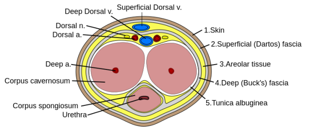
- Layers surrounding the corpora cavernosa, from superficial to deep (5):
- Skin
- Dartos (continuous with Scarpa’s and Colle’s fasciae)
- Tela subfascialis (very thin connective tissue layer)
- Buck’s fascia
- Tunica albuginea

Corpus cavernosum[edit | edit source]
- The erectile bodies; comprised of 2 spongy, paired cylinders
- Contained in the tunica albuginea
- The erectile tissues of the normal corpora cavernosa are separated from the tunica by the space of Smith
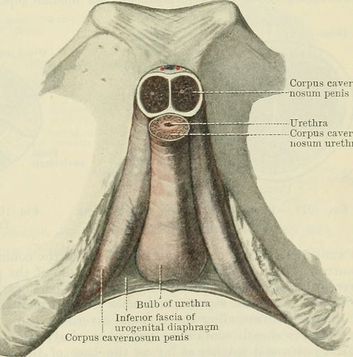
- Proximal ends, the crura, originate at the undersurface of the puboischial rami as separate structures but merge under the pubis to form the body of the penis and remain attached up to the glans
- A septum separates the corpora cavernosa but is permeable distally to allow for free communication between their vascular spaces

- Each corpus cavernosum is a conglomeration of sinusoids, larger in the center and smaller in the periphery.
- In the flaccid state, the blood slowly diffuses from the central to the peripheral sinusoids, and the blood gas levels are similar to those of venous blood.
- During erection, the rapid entry of arterial blood to both the central and the peripheral sinusoids changes the intracavernous blood gas levels to those of arterial blood
Corpus spongiousum[edit | edit source]
- The urethra travels through the corpus spongiosum, with its proximal segment known as the bulb.
- The glans penis is an expansion of the corpus spongiosum
Tunica albuginea[edit | edit source]
- Tough connective tissue layer composed primarily of type I collagen
- A bilayered structure (inner circular, outer longitudinal) with multiple sublayers.
- Outer longitudinal layer
- Completely absent on the ventral groove between the 5 and 7 o'clock positions.
- The most vulnerable area in the tunica albuginea
- Most prostheses tend to extrude here
- The lack of tunica albuginea on the ventral groove may contribute to greater ease of dorsal buckling and explain why most PD patients exhibit dorsal curvature
- 60-70% of plaques are located on the dorsal aspect of the penis and are usually associated with the septum
- The most vulnerable area in the tunica albuginea
- Completely absent on the ventral groove between the 5 and 7 o'clock positions.
- Outer longitudinal layer
- Covers the corpora cavernosa and *spongiosum
- *The corpus spongiosum lacks an outer longitudinal layer or intracorporeal struts, ensuring a low-pressure structure during erections
Buck fascia[edit | edit source]
- External to the tunica albuginea
- Surrounds corpora cavernosae; ventrally, splits to surround the corpus spongiosum
- Distally, fuses with the glans at corona
- The paired dorsal arteries, paired dorsal nerves, and deep dorsal vein are deep to Buck’s fascia
- The superficial dorsal penile vein is external to Buck’s fascia
- A penile fracture is a tear in the tunica albuginea; the hematoma is contained by Buck’s fascia
Penile skin[edit | edit source]
- Very elastic
- Only glandular elements are the smegma-producing glands, called glands of Tyson, located at the base of the corona
External penile support[edit | edit source]
- Consists of 2 ligamentous structures: the fundiform and suspensory ligaments
- The fundiform ligament arises from Colles fascia and is lateral, superficial, and not adherent to the tunica albuginea of the corpora cavernosa.
- The suspensory ligament arises from Buck fascia and consists of two lateral bundles and one median bundle
Vasculature[edit | edit source]
Arterial supply[edit | edit source]
- Deep arterial system:
- The internal pudendal artery, (derived from the anterior branch of the internal iliac) gives off the perineal branch and posterior scrotal arteries before continuing on to become the common penile artery.
- The common penile artery has 3 paired branches: CBD
- Cavernosal artery
- Penetrates the corpus cavernosum in the penile hilum to nearly the center of the erectile tissue
- Provides straight and helicine arteries that supply the cavernous sinuses; the helicine arteries supply the trabecular erectile tissue and the sinusoids. These helicine arteries are contracted and tortuous in the flaccid state and become dilated and straight during erection
- Effects tumescence of the corpus cavernosum
- Bulbourethral artery
- Supplies the corpus spongiosum, urethra, and glans
- Penetrates the perineal membrane where it enters the corpus spongiosum from above at its posterolateral border
- Dorsal artery
- Supplies the dorsal surfaces of the corporeal bodies and circumferential branches to the urethra and the corpus spongiosum; responsible for engorgement of the glans during erection; supplies distal shaft skin
- Travels between the crus and the pubis then between the dorsal vein and the dorsal penile nerve, all of which attach to the underside of Buck fascia, then travels distally toward the glans
- Distally, these arteries join to form a vascular ring near the glans.
- Can be a great deal of variability in penile arteries.
- The bulbourethral [“and urethral”?] arteries are situated outside the tunica albuginea of the corpus spongiosum on the lateral and dorsal sides
- The cavernous artery and branches of the dorsal artery that supply the corpus cavernosae are surrounded by a periarterial soft-tissue sheath, which protects the arteries from occlusion by the tunica albuginea during erection
- Accessory penile arteries
- Exist in many instances
- May constitute the dominant or only arterial supply to the corpus cavernosum in some males.
- Accessory pudendal arteries arise most commonly from the obturator artery.
- Cavernosal artery
- Superficial arterial system:
- Branches from the external pudendal (originates from femoral artery) artery supply the penile skin; these vessels run in the dartos fascia layer.
- The blood supply of the skin is independent of the erectile bodies. The rich anastomotic network makes penile skin ideal for mobilization on a vascular pedicle
Venous drainage[edit | edit source]
- Drainage from the 3 corpora originates in tiny venules leading from the peripheral sinusoids immediately beneath the tunica albuginea. These venules travel in the trabeculae between the tunica and the peripheral sinusoids to form the subtunical venous plexus before exiting as the emissary veins
- Deep dorsal vein
- Main venous drainage of the glans penis and distal two thirds of the corpora cavernosa
- Usually a single vein, but sometimes more than one deep dorsal vein exists
- Beneath Buck fascia and runs between the paired dorsal arteries (see above)
- Travels between the corporal bodies and runs upward behind the symphysis pubis, between the inferior pubic arch and the striated urinary sphincter, to join the periprostatic venous plexus
- Trifurcates into
- Central superficial branch
- Pierces endopelvic fascia between puboprostatics
- Drains retropubic fat, anterior bladder and anterior prostate
- Outside prostatic fascia
- Drains into the internal pudendal vein
- 2 lateral plexuses
- Sweep on lateral surface of prostate
- Drain prostate and rectum; these then communicate with vesical plexuses on lower part of bladder
- 3-5 inferior vesical veins emerge from these lateral vesical plexuses and drain into the internal iliac vein
- In females, the dorsal clitoral vein bifurcates to empty into lateral vaginal plexuses; these connect with vesical, uterine, ovarian, and rectal plexuses to drain into the internal iliac vein
- Connections exist between pelvic plexuses and emissary veins of pelvic bones and vertebral plexuses; proposed route for dissemination of pelvic processes to pelvic & axial bone
- The veins of the three systems communicate variably with each other.
- Central superficial branch
- Superficial dorsal vein
- External to Buck fascia
- Drains into saphenous vein
Lymphatic supply[edit | edit source]
- Penile shaft lymphatics converge on the dorsum and ramify to both sides of the groin to drain into the superficial and deep inguinal lymph nodes
Innervation[edit | edit source]
Somatic[edit | edit source]
- Paired dorsal nerves of the penis (paired, S2-S4)
- Arises in the Alcock canal as the first branch of the pudendal nerve (S2-4)
- Runs over the surface of the obturator internus under the levator, runs deep to the urogenital diaphragm, and passes through the deep transverse perineal muscle to run along the dorsum of the penis accompanied by the dorsal vein and dorsal artery.
- Supplies sensory innervation to the glans
- Small branches of the perineal nerve supply the ventrum of the penis as distally as the glans
- The skin of the penis is innervated by branches of the genitofemoral nerve
Autonomic[edit | edit source]
- Paired cavernous nerves (S2-4)
- Arise from the pelvic plexus from the lateral surface of the rectum
- Run posterolateral to the apex, mid-portion and base of the prostate anterior to Denonvilliers’ fascia between the posterolateral surface of the prostate and the rectum to lie between the lateral pelvic fascia and the prostatic fascia. The branches from the cavernous nerve accompany the branches of the prostatovesicular artery and provide a macroscopic landmark for nerve-sparing radical prostatectomy i.e. neurovascular bundle.
- Innervates the corpus cavernosum and penile urethra
Questions[edit | edit source]
- What explains the lack of rigidity of the corpus spongiosum during an erection?
- What is the relationship of the superficial dorsal penile vein to Buck’s fascia? Dorsal artery? Deep dorsal vein?
- How many dorsal arteries and deep dorsal veins are there?
- What is the arterial blood supply to the penis and what is the origin of each artery?
- Which artery is responsible for glans engorgement during erection?
Answers[edit | edit source]
- What explains the lack of rigidity of the corpus spongiosum during an erection?
- What is the relationship of the superficial dorsal penile vein to Buck’s fascia? Dorsal artery? Deep dorsal vein?
- Superficial dorsal vein is superficial to Buck’s fascia
- Dorsal artery and deep dorsal vein are deep to Buck’s fascia
- How many dorsal arteries and deep dorsal veins are there?
- Two dorsal arteries, single dorsal vein
- What is the arterial blood supply to the penis and what is the origin of each artery?
- The common penile artery gives off 3 branches that supply the penis including the cavernosal artery, bulbourethral artery, and dorsal artery
- Which artery is responsible for glans engorgement during erection?
- Dorsal artery
References[edit | edit source]
- Wein AJ, Kavoussi LR, Partin AW, Peters CA (eds): CAMPBELL-WALSH UROLOGY, ed 11. Philadelphia, Elsevier, 2015, chap 21