Stones: Diagnosis and Evaluation
Jump to navigation
Jump to search
See Original 2019 AUA Evaluation and Medical Management of Stones Guidelines
See 2019 AUA Evaluation and Medical Management of Stones Guideline Notes
See 2016 CUA Evaluation and Medical Management of Stones Guideline Notes
Imaging for Stone Disease[edit | edit source]
Plain Abdominal Film[edit | edit source]
- Findings
- Radiolucent stones (6):
- Uric acid
- Matrix
- Medication stones (4):
- Xanthine
- Triamterene
- 2,8-dihydroxyadenine
- Indinavir
- Radioopaque stones (4):
- Calcium oxalate
- Calcium phosphate
- Poorly radioopaque:
- Magnesium ammonium phosphate (struvite)
- Cystine stones
- Although magnesium ammonium phosphate and cystine stones are often radioopaque, they are not as dense as calcium oxalate or calcium phosphate stones
- Nephrocalcinosis
- Formation of diffuse deposits of calcium throughout the kidneys
- Usually occurs within the renal medulla but occasionally it has been found in the cortex or within both the medulla and the cortex
- Minute calcifications seen in early stages may not be visible
- Can give rise to renal colic and hydronephrosis from dislodged calcific foci
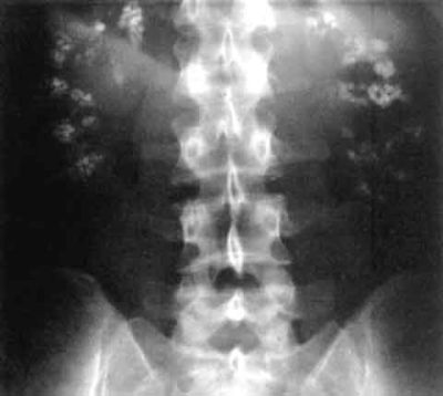 Plain film x-ray demonstrating bilateral diffuse calcium deposits in the kidneys. Source: Wikipedia
 - Causes§
- Medulla
- Type 1 (distal) RTA
- Hyperparathyroidism
- Medullary sponge kidney
- Hypervitaminosis D
- Milk-alkali syndrome
- Sarcoidosis
- Hyper/hypothyroidism
- Other pathological hypercalcemic or hypercalciuric states
- Cushing syndrome
- Multiple myeloma
- Bartter syndrome
- Bone metastases
- Pyramids
- Hyperuricemia
- Infection (particularly renal tuberculosis)
- Sickle cell disease (leading to infarction and subsequent dystrophic calcification)
- Renal papillary necrosis
- Drugs
- Furosemide abuse
- Corticol COAG
- Corticol necrosis
- Oxalosis (Primary hyperoxaluria)
- Alport syndrome
- Glomerulonephritis (chronic)
- Medulla
- Formation of diffuse deposits of calcium throughout the kidneys
- Radiolucent stones (6):
- Test characteristics§
- Sensitivity: 57%
- Specificity: 76%
- Advantage(s)
- Availability
- Relatively low radiation exposure
- 0.7 mSv with KUB§
- Cost (least expensive)
- Disadvantages
- Inability to visualize small stones
- Inability to visualize stones due to overlying/underlying anatomy (bones, phleboliths, etc.)
- Underestimates >90% of stones >10mm

Ultrasound[edit | edit source]
- Test characteristics§
- Sensitivity: 84%
- Specificity: 53%
- Advantages
- Availability
- No radiation exposure
- Cost (5x cost of KUB)§
- Disadvantages (2):
- Inability to visualize most ureteral stones
- Poor correlation between measured and actual stone size and location
- If stone
- <10mm, US underestimates size of stone 1/3 of the time
- >10mm, US overestimates size of stone 1/3 of the time
- US and CT measurements correlate 2/3 of the time
- If stone
CT scan (without contrast)[edit | edit source]
- Findings
- Pure uric acid stones have much lower Hounsfield units than calcium types
- Forniceal extravasation
- Usually associated with a small distal ureteral calculus.
- Should be similarly to other ureteral stones: intervention should be undertaken when there is an associated fever, nausea/vomiting, or unrelenting pain. Otherwise, conservative observation is appropriate.
- Test characteristics§
- Sensitivity: 95%
- Specificity: 98%
- Advantages
- Most sensitive modality for stones
- Disadvantages
MRI[edit | edit source]
- Test characteristics§
- Sensitivity: 82%
- Specificity: 98%
- Findings
- Stones appear as filling defects
- Advantages
- No exposure to radiation
- Disadvantages
- Availability
- Cost (most expensive, 30x cost of KUB)§
Acute Diagnosis and Management[edit | edit source]
- Diagnosis and Evaluation
- History and Physical Exam
- History
- New-onset urgency and frequency may indicate a stone at the UVJ irritating the bladder
- Sudden relief of flank pain may indicate either passage or forniceal rupture as the pressure in the collecting system dramatically decreases.
- Physical Exam
- Abdomen
- Costovertebral angle tenderness
- History
- Labs
- Urinalysis +/- culture
- CBC
- Serum creatinine
- Imaging
- History and Physical Exam
- Management
- Renal colic pain management[1]
- Toradol 30 mg IV
- Cardiac Lidocaine 1.5 mg/kg IV in 100 mL NS over 10 minutes (MAX 200 mg)
- Acetaminophen 1000 mg PO
- 1 L 0.9% NS bolus
- If obstructing stones with suspected infection, must urgently drain the collecting system with a stent or nephrostomy tube and delay stone treatment★
- Definitive management of the stone should not be undertaken until sepsis has resolved and the infection has been treated with an appropriate course of antibiotic therapy.
- Renal colic pain management[1]
Diagnosis and Evaluation of Metabolic Stone Disease[edit | edit source]
UrologySchool.com Summary[edit | edit source]
AUA★[edit | edit source]
- Patients with newly diagnosed kidney or ureteral stones should undergo a sScreening Evaluation evaluation consisting of
- History and Physical Exam
- Laboratory (5)
- Urinalysis +/- culture
- Serum electrolytes (Na, K, Cl, HCO3)
- Serum calcium
- Serum creatinine
- Serum uric acid
- Imaging
- Obtain or review available imaging studies to quantify stone burden.
- Extended evaluation
- One or two 24-hour urine collections
- Indications (7):
- Recurrent stone formers
- Family history of stone disease
- Solitary kidney
- Malabsorptive intestinal disease or resection
- Obesity
- Recurrent UTIs
- Medical conditions predisposing to stones (e.g., RTA Type 1, primary hyperparathyroidism, gout, diabetes mellitus type)
- Indications (7):
- One or two 24-hour urine collections
Goals of Evaluation[edit | edit source]
- Identify potential associated metabolic disorders such as (5)
- Distal renal tubular acidosis (RTA)
- Primary hyperparathyroidism
- Enteric hyperoxaluria
- Cystinuria
- Gouty diathesis
- Reduce risk of stone recurrence
- First-time stone formers have been estimated to have a 50% risk for recurrence within the subsequent 10 years
- Patients at higher risk for repeat episodes (6):
- Family history of stones
- Intestinal disease (particularly when causing chronic diarrheal states)
- Pathologic skeletal fractures
- Osteoporosis
- UTI
- Gout
History and Physical Exam[edit | edit source]
History[edit | edit source]
- Signs and Symptoms
- Flank pain
- Hematuria
- Lower urinary tract symptoms
- Risk factors
- Conditions associated with stone disease (8):
- Obesity
- Hyperthyroidism
- Gout
- Renal tubular acidosis (RTA) type 1
- Diabetes mellitus type 2
- Bone disease
- Primary hyperparathyroidism
- Malabsorptive gastrointestinal states due to bowel resection, bariatric surgery or bowel or pancreatic disease
- Chronic diarrhea that could be caused by inflammatory bowel disease (Crohn disease, ulcerative colitis) or irritable bowel syndrome
- Gout may predispose the patient to hyperuricosuria or gouty diathesis with either uric acid calculi or calcium oxalate stone formers
- Surgical history should be obtained focusing particularly on bariatric surgery and surgeries of the intestinal tract.
- Roux-en-Y-gastric bypass surgery may significantly increase the overall risk for stone formation
- In contrast to gastric bypass surgery, restrictive bariatric surgeries such as gastric sleeve or gastric band do not seem to increase the risk for kidney stones
- Dietary history
- Should include average daily intake of fluids (amount and specific beverages), protein (types and amounts), calcium, sodium, high oxalate-containing foods, fruits and vegetables and over-the-counter supplements.
- Nutritional factors associated with stone disease, depending on stone type and risk factors, include
- Calcium intake below or significantly above the recommended dietary allowance (RDA)
- Low fluid intake
- High sodium intake
- Limited intake of fruits and vegetables
- High intake of animal-derived purines
- Nutritional factors associated with stone disease, depending on stone type and risk factors, include
- Should include average daily intake of fluids (amount and specific beverages), protein (types and amounts), calcium, sodium, high oxalate-containing foods, fruits and vegetables and over-the-counter supplements.
- Medications
- Stone-provoking medications or supplements (9):
- Triamterene
- Carbonic anhydrase inhibitors such as topiramate, acetazolamide, zonisamide
- Probenecid
- Some protease inhibitors
- Lipase inhibitors
- Chemotherapy
- Vitamin C
- Vitamin D
- Calcium
- Stone-provoking medications or supplements (9):
- Conditions associated with stone disease (8):
Physical Exam[edit | edit source]
- General
- Body mass index
- Increased BMI, larger waist size, and weight gain are correlated with an increased risk for stone episodes
- The association of obesity and uric acid stone formation is primarily due to change in urinary pH
- The association of obesity with calcium oxalate stone formation is primarily due to increased excretion of promoters of stone formation (oxalate, uric acid, sodium, and phosphorus)
- Increased BMI, larger waist size, and weight gain are correlated with an increased risk for stone episodes
- Body mass index
- Flank
- Costovertebral tenderness
Laboratory[edit | edit source]
Urinalysis +/- culture +/- microscopy[edit | edit source]
- Urinalysis should include pH
- Normal pH should be between 5.8-6.2§
- pH > 7.0 is suggestive of infection lithiasis or RTA
- pH < 5.5 suggests uric acid lithiasis secondary to gouty diathesis
- Normal pH should be between 5.8-6.2§
- Urine culture
- Should be obtained in patients with a urinalysis suggestive of UTI.
- Presence of urea-splitting organisms, such as Proteus species, raises the possibility of struvite stones
- Many infected calculi will harbor bacteria even after treatment with broad-spectrum antibiotics
- Half of infected calculi grow bacterial cultures that are different from the preoperative urine specimen
- Urine microscopy
- Identify crystals pathognomonic of stone type.
Serum chemistries[edit | edit source]
- Includes
- Electrolytes (Na, K, Cl, HCO3)
- Calcium
- Uric acid
- Creatinine
- May suggest underlying medical conditions associated with stone disease (e.g., primary hyperparathyroidism, gout, RTA type 1 or other metabolic derangements)
- Assessment of underlying renal function is necessary
Parathyroid hormone (PTH)[edit | edit source]
- Indicated as part of the screening evaluation if primary hyperparathyroidism is suspected
- Primary hyperparathyroidism should be suspected when (3):
- Mid-range PTH despite high or high normal serum calcium
- Increased urinary calcium
- Predominantly calcium phosphate stone composition
- Primary hyperparathyroidism should be suspected when (3):
- Measurement of vitamin D levels may be helpful as low vitamin D levels may mask primary hyperparathyroidism, or contribute to secondary hyperparathyroidism.
- A high or high normal intact PTH in these settings should prompt further endocrine evaluation, imaging or referral for consideration of parathyroidectomy.
Stone composition, if available[edit | edit source]
- When a stone is available, a stone analysis should be obtained at least once.
- Can direct metabolic investigation or potentially obviate the need for a complete metabolic evaluation
- Calcium phosphate stone composition associated with:
- RTA Type 1
- Primary hyperparathyroidism
- Medullary sponge kidney
- Use of carbonic anhydrase inhibitors
Imaging[edit | edit source]
- Obtain or review available imaging studies to quantify stone burden. ★
- No standard definition exists for complete and partial staghorn stones
- Most consider complete staghorn stones to occupy the entire renal collecting system, whereas partial staghorn stones occupy less.
Metabolic/Extended Diagnostic Evaluation[edit | edit source]
- Consists of one or two 24-hour urine collections obtained on a random diet★
Indications[edit | edit source]
- AUA (7):★
- Recurrent stone formers
- Family history of stone disease
- Solitary kidney
- Malabsorptive intestinal disease or resection
- Obesity
- Recurrent UTIs
- Medical conditions predisposing to stones (e.g., RTA Type 1, primary hyperparathyroidism, gout, diabetes mellitus type)
- Included in other lists
- Pathological skeletal fractures
- Osteoporosis
- Infirm health (unable to tolerate repeat stone episodes)
- Anatomic abnormalities
- Stones composed of cystine, uric acid, and struvite
- Children should generally be evaluated because of concerns about renal damage and long-term sequelae of stone recurrence
24-hour urine collections[edit | edit source]
- Can be used to inform and monitor treatment protocols
- Analyzed at minimum for (9): ★
- Volume
- pH
- Creatinine
- Sodium
- Potassium
- Calcium
- Oxalate
- Uric acid
- Citrate
- In stone formers with known cystine stones or a family history of cystinuria or for those in whom cystinuria is suspected, urinary cystine should additionally be measured.
- Sulfate can be added to assess the volume of protein loading from animal meat
- The accuracy of a 24-hour urine collection should be assessed prior to interpretation of results.
- To assess the adequacy of collection, 24-hour urinary creatinine excretion should be evaluated, taking into account patient gender and body weight, as well as patient recall of the start and end times of his or her collection, should be considered
- Significant aberrations in total creatinine excretion from estimated volumes (males 20-25mg/kg and females 15-20mg/kg in 24 hours) imply incomplete collection, overcollection, greater than expected muscle mass, or less than expected muscle mass
- For abnormally collected 24 hour urine collections, can divide metabolite excretion by creatinine excretion to compare collections
- Significant aberrations in total creatinine excretion from estimated volumes (males 20-25mg/kg and females 15-20mg/kg in 24 hours) imply incomplete collection, overcollection, greater than expected muscle mass, or less than expected muscle mass
- To assess the adequacy of collection, 24-hour urinary creatinine excretion should be evaluated, taking into account patient gender and body weight, as well as patient recall of the start and end times of his or her collection, should be considered
- Markers of protein intake, such as urine urea nitrogen or urinary sulfate, are reflective of animal protein intake and can be used to assess dietary adherence.
- Urinary potassium measured at baseline can be compared to urinary potassium obtained during follow-up to gauge compliance with medication regimens.
- Primary hyperoxaluria should be suspected when urinary oxalate excretion > 75 mg/day in adults without bowel dysfunction. These patients should be considered for referral for genetic testing and/or specialized urine testing
- Fast and calcium load testing should not be performed routinely to distinguish among types of hypercalciuria
- If a patient with calcium urolithiasis uses calcium supplements, 24-hour urine samples should be collected on and off the supplement.
- If urinary supersaturation of the calcium salt in question increases during the period of supplement use, the supplement should be discontinued.
Questions[edit | edit source]
- What is the risk of stone recurrence at 10 years in first-time stone formers?
- What is the microscopic appearance of common urinary calculi?
Answers[edit | edit source]
1. 50%
Next Chapter: Diet and Pharmacologic Management[edit | edit source]
References[edit | edit source]
- Wein AJ, Kavoussi LR, Partin AW, Peters CA (eds): CAMPBELL-WALSH UROLOGY, ed 11. Philadelphia, Elsevier, 2015, chap 52
- Pearle, Margaret S., et al. "Medical management of kidney stones: AUA guideline." The Journal of urology 192.2 (2014): 316-324.