Uncategorized files
Jump to navigation
Jump to search
Showing below up to 48 results in range #1 to #48.
View (previous 50 | next 50) (20 | 50 | 100 | 250 | 500)
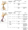 1000px-1810 Major Pituitary Hormones.jpg 1,000 × 1,058; 129 KB
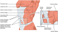 1112 Muscles of the Abdomen Anterolateral.png 1,046 × 578; 450 KB
 1818 The Adrenal Glands.jpg 1,102 × 316; 250 KB
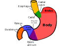 2560px-Regions of stomach.svg.png 2,560 × 1,975; 197 KB
 2560px-Stomach blood supply.svg.png 2,560 × 856; 192 KB
 43414588252 df2480a453 o.jpg 851 × 746; 148 KB
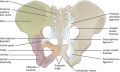 817 Ligaments of Pelvis.jpg 873 × 523; 222 KB
 Abdotrauma.png 1,018 × 782; 434 KB
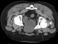
 Artificial urethral sphincter - CT axial 001.jpg 3,068 × 1,170; 307 KB
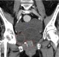 Artificial urethral sphincter - CT coronar 001.jpg 1,422 × 1,365; 178 KB
 Bladder Diverticulum.jpg 1,161 × 894; 379 KB
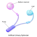 Blausen 0059 ArtificialUrinarySphincter.png 1,500 × 1,500; 471 KB
 CT colovesical fistila.jpg 781 × 807; 122 KB
 Common Sites of Lower Abdominal Hernias.jpg 463 × 402; 88 KB
 Cystoscopy - Uretereal Cancer.jpg 768 × 576; 105 KB
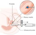 Diagram showing a transperineal prostate biopsy CRUK 473.jpg 877 × 862; 114 KB
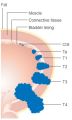 Diagram showing the T stages of bladder cancer CRUK 372.jpg 243 × 405; 25 KB
 Dolbeau technique of perineal lithotomy.jpg 512 × 523; 82 KB
 Femoral triangle.png 550 × 600; 134 KB
 Figure 28 01 06.jpg 781 × 718; 295 KB
 Gray 1913 1285.png 2,400 × 2,107; 4.83 MB
 Hypercortisolism.jpg 1,333 × 750; 153 KB
 Hypothalamic and pituitary hormones.png 512 × 275; 64 KB
 Hypothalamic hormones.png 512 × 275; 64 KB
 Hypothalamus-Hypophysis-Testicle-Hormone-Axis.svg.png 512 × 321; 19 KB
 Inguinal fossae.png 1,059 × 534; 111 KB
 Kidney CT Mass Axial.png 806 × 545; 287 KB
 Kidney CT Mass Sagittal.png 807 × 908; 312 KB
 Laparoscopic view of inguinal region.jpg 640 × 480; 155 KB
 Leftrenalarteryinjury.png 1,173 × 978; 834 KB
 Male Infertility.png 1,079 × 1,316; 139 KB
 Nephrocalcinosis.jpg 480 × 430; 9 KB
 Neuroblastoma 103.jpg 600 × 600; 51 KB
 Penis Anatomy Cross Section.png 764 × 328; 69 KB
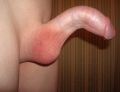 Peyronie's disease.jpg 2,993 × 2,304; 2.27 MB
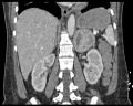 Phaeochromozytoma CT coronal.jpg 512 × 410; 107 KB
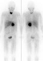 Pheochromocytoma Scan.jpg 526 × 747; 244 KB
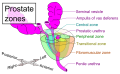 Prostate zones.png 1,231 × 754; 207 KB
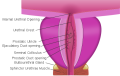 Prostatic urethra .png 512 × 326; 73 KB
 Renal parenchymal phase CT of transitional cell carcinoma.jpg 937 × 307; 99 KB
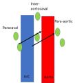 Retroperitoneal lymph flow.jpg 448 × 495; 28 KB
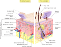 Skin layers.png 1,282 × 997; 621 KB
 Spermatogenesis Diagram.jpg.png 640 × 480; 154 KB
 Spermatozoa anatomy.png 1,000 × 911; 260 KB
 Stellate scar in right renal mass.jpg 500 × 404; 69 KB
 Steroidogenesis.png 512 × 321; 47 KB
 TAJU A 1589748 F0005 OC.jpg 800 × 903; 27 KB
 Wiki Penis Base Cross Section.jpg 1,014 × 1,024; 185 KB


