Female Urethral Diverticulae
Jump to navigation
Jump to search
Background[edit | edit source]
- Definition of female urethral diverticulum: a variably sized urine-filled periurethral cystic structure adjacent to the urethra within the confines of the pelvic fascia, connected to the urethra via an ostium
Epidemiology[edit | edit source]
- Prevalence: unknown; reported in up to 1-6% of adult females in some series
- Majority of patients present between the 3rd-7th decade of life
Classification[edit | edit source]
- Classified: congenital vs. acquired
- Vast majority are acquired and diagnosed in adult females; few are congenital
Risk factors[edit | edit source]
- Acquired
- Most acquired female UD are thought to originate from pathologic processes, usually infection, involving the periurethral [Skene] glands
Anatomy[edit | edit source]
- Size of diverticulum can be few mm to several cm
- Ostium of the urethral diverticulum is located postero/ventrolaterally at the 4 and 8 o’clock positions in the mid- or distal urethra in >90%, corresponding to the location of the periurethral glands
- The interior surface of the UD may be urothelial, squamous, columnar, or cuboidal epithelium, or mixed. In some cases, the epithelium is absent and the wall of the UD consists of only fibrous tissue.
- 2/3 of resected UD demonstrate inflammatory changes. Most UD demonstrate benign histopathology but premalignant and malignant changes can be seen.
- Most common malignant histology in urethral diverticulum is adenocarcinoma
- Recall, most common malignant histology in females urethral carcinoma is squamous cell carcinoma while in male urethral cancers most common histology is urothelial; see Urethral Tumours Chapter Notes)
- Most common malignant histology in urethral diverticulum is adenocarcinoma
Differential Diagnosis[edit | edit source]
- Periurethral Masses Other Than Urethral Diverticula (7):
- Periurethral Bulking Agents
- Vaginal Leiomyoma
- Skene Gland Abnormalities
- Gartner Duct Abnormalities
- Vaginal Wall Cysts
- Urethral Mucosal Prolapse
- Urethral Caruncle
Urethral Caruncle[edit | edit source]
- An inflammatory lesion of the distal urethra
- Epidemiology
- Most commonly diagnosed in postmenopausal women
- Diagnosis and Evaluation
- Physical Exam
- Urethral prolapse is similar in appearance, but is circumferential while caruncles are focal
- Physical Exam
- Management
- Most recommend initial conservative management with topical estrogen or anti-inflammatory creams and sitz baths.
- Large or refractory lesions may be managed with simple excision.
- If the lesion is atypical in appearance or behavior, excision may be warranted to exclude other entities.
Diagnosis and Evaluation[edit | edit source]
UrologySchool.com Summary[edit | edit source]
- History and Physical Exam
- Labs
- U/A +/- culture
- Imaging
- Options (5)
- Double-balloon positive pressure urethrography
- VCUG
- Intravenous urography
- US
- MRI
- Options (5)
- Other
- Cystourethroscopy
- +/- UDS
History and Physical Exam[edit | edit source]
History[edit | edit source]
- Signs and Symptoms
- Notable for a range of clinical presentations ranging from completely asymptomatic (up to 20% of patients), incidentally noted lesions on physical examination or imaging, to very debilitating, painful vaginal masses associated with incontinence, stones, severe dyspareunia, and/or tumors
- Most common symptoms (3):
- Storage LUTS
- Pain
- Infection
- Multiple bouts of recurrent cystitis should alert the possibility of a urethral diverticulum
- Reinfection, inflammation, and recurrent obstruction of the neck of the cavity are theorized to result in patient symptoms and enlargement of the diverticulum. This expansion occurs most commonly ventrally, resulting in the classic anterior vaginal wall mass palpated on physical examination in some patients with UD. However, it is important to note that these may also expand laterally, or even dorsally, about the urethra. Eventually, the abscess cavity ruptures into the urethral lumen, resulting in the communication between the UD and the urethral lumen.
- Multiple bouts of recurrent cystitis should alert the possibility of a urethral diverticulum
- Other symptoms (8):
- Dysuria
- Hematuria
- Post-void dribbling
- Urinary retention
- Incontinence (stress or urge)
- Dyspareunia
- Vaginal mass
- Patients may present with complaints of a tender or nontender anterior vaginal wall mass, which upon gentle compression may reveal retained urine or purulent discharge per the urethral meatus.
- Vaginal discharge
- Vaginal pruritis is not a symptom associated with urethral diverticulum
- Size of the UD does not correlate with symptoms
Physical Exam[edit | edit source]
- Genitals
- Urethra may be gently “stripped” or “milked” distally in an attempt to express purulent material or urine from within the urethral diverticulum
Laboratory[edit | edit source]
- Urinalysis +/- culture

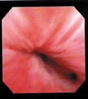
Cystoscopy with visualization of urethral diverticulum ostium at the 4 o’clock position just proximal to the mid-urethral sphincter. Source
Imaging[edit | edit source]
- No single study can be considered the gold standard
- Options (5)
- Double-balloon positive pressure urethrography
- VCUG
- Intravenous urography
- US
- MRI
- Imaging cannot reliably diagnose malignancy in a urethral diverticulum
Other[edit | edit source]
- Cystourethroscopy
- Patients with UD are often highly symptomatic, and endoscopic examination can be very difficult to initiate or complete without anesthesia.
- The UD ostium can be very difficult to identify in some patients
- Stones within the urethral diverticulum may be diagnosed 4-10% of the time
Management: observation vs. intervention[edit | edit source]
Options[edit | edit source]
- Observation
- Intervention
- Surgical interventions (5):
- Excision with reconstruction (most common surgical approach)
- Marsupialization (transurethral and open)
- Endoscopic unroofing
- Fulguration
- Incision and obliteration with oxidized cellulose or polytetrafluoroethylene
- Surgical interventions (5):
Observation[edit | edit source]
- Very little is known regarding the natural history of untreated UD. For these reasons, and because of the lack of symptoms in selected cases, some patients may not desire surgical therapy.
- There are reports of malignancy arising in UD. Therefore, patients should be counselled on the risk of malignancy with nonoperative management
- Patients electing nonoperative management can be treated with low-dose antibacterial suppressants and digital stripping of the anterior vaginal wall following micturition to prevent postvoid dribbling and reduce the risk of UTI resulting from stasis in the UD.
- Whether long-term surveillance is required in these patients, with periodic physical examinations, radiographic imaging, or endoscopic examination, is unknown.
Intervention[edit | edit source]
Indications[edit | edit source]
- Symptomatic patients, including those with dysuria, dyspareunia, refractory bothersome postvoid dribbling, recurrent UTIs, and pelvic pain, may be offered surgical excision.
Options (5):[edit | edit source]
- Excision (urethral diverticulectomy) with reconstruction (most common surgical approach)
- Marsupialization (transurethral and open)
- Endoscopic unroofing
- Fulguration
- Incision and obliteration with oxidized cellulose or polytetrafluoroethylene
Urethral Diverticulectomy[edit | edit source]
- Principles (8):
- Mobilization of a well-vascularized anterior vaginal wall flap(s)
- Preservation of the periurethral fascia
- Identification and excision of the neck, or ostium, of the UD
- Removal of entire UD wall or sac (mucosa)
- Watertight urethral closure
- Multilayered, nonoverlapping closure with absorbable suture
- Closure of dead space
- Preservation or creation of continence
- The location and competence of the urethral sphincters have important implications when considering surgical repair of urethral diverticulectomy because of the anatomic overlap of these two entities.
- Varying degrees of sphincteric compromise may exist prior to intervention because of the location of diverticulum relative to the proximal and distal urinary sphincter mechanisms, or sphincteric compromise may coexist with UD as a result of other factors.
- Technique
- Successful excision of a urethral diverticulum involves removal of the ostium that connects with the urethral lumen. This often results in direct visualization of the urethral catheter within the urethral lumen during surgery. The urethral defect is closed primarily with absorbable suture in a watertight fashion following completion of the removal of the sac.
- Additional procedures such as buccal mucosal urethroplasty, Martius flap, or vaginal flaps are not necessary to close the urethra.
- Synthetic materials (e.g., mid-urethral polypropylene mesh) should not be used in an anti-incontinence procedure synchronously with urethral diverticulum surgery because of the potentially increased risk of urethral erosion and infection
- Successful excision of a urethral diverticulum involves removal of the ostium that connects with the urethral lumen. This often results in direct visualization of the urethral catheter within the urethral lumen during surgery. The urethral defect is closed primarily with absorbable suture in a watertight fashion following completion of the removal of the sac.
- Adverse Events
- Recurrent UTIs
- Urinary incontinence
- Recurrent urethral diverticulum
- Urethrovaginal fistula (uncommon)
- Size of diverticulum does correlated with risk of recurrence following surgical repair
- Postoperatively, some patients will have persistence or recurrence of their preoperative symptoms.
Questions[edit | edit source]
- How do female urethral diverticuli usually develop?
- Where is the ostium of most female urethral diverticuli located?
- What is the most common histology of malignancy in a female urethral diverticulum? Female urethral cancer? Male urethral cancer?
- List signs and symptoms associated with a female urethral diverticulum?
- What is the lymphatic drainage of the female urethra?
- What is the lymphatic drainage of the male urethra?
- Differential diagnosis of periurethral mass?
- What is the most common surgical approach to treat a female urethral diverticulum?
Answers[edit | edit source]
- How do female urethral diverticuli usually develop?
- Infection involving the periurethral glands
- Where is the ostium of most female urethral diverticuli located?
- Posterolaterally in the mid- or distal urethra
- What is the most common histology of malignancy in a female urethral diverticulum? Female urethral cancer? Male urethral cancer?
- Female urethral diverticulum: adenocarcinoma
- Female urethral cancer: squamous
- Male urethral cancer: urothelial
- List signs and symptoms associated with a female urethral diverticulum?
- Storage LUTS, pain, infection, dysuria, dyspareunia, dribbling, vaginal mass, hematuria, vaginal discharge, voiding symptoms, retention,
- What is the lymphatic drainage of the female urethra?
- Proximal: external iliac nodes
- Distal: superficial inguinal nodes
- What is the lymphatic drainage of the male urethra?
- Posterior: pelvic nodes
- Anterior: superficial inguinal lymph nodes
- Differential diagnosis of periurethral mass?
- Vaginal Leiomyoma
- Skene Gland Abnormalities
- Gartner Duct Abnormalities
- Vaginal Wall Cysts
- Urethral Mucosal Prolapse
- Urethral Caruncle
- Periurethral Bulking Agents
- What is the most common surgical approach to treat a female urethral diverticulum?
- Excision and reconstruction
References[edit | edit source]
- Wein AJ, Kavoussi LR, Partin AW, Peters CA (eds): CAMPBELL-WALSH UROLOGY, ed 11. Philadelphia, Elsevier, 2015, chap 90
- Greiman, Alyssa K., Jennifer Rolef, and Eric S. Rovner. "Urethral diverticulum: a systematic review." Arab journal of urology 17.1 (2019): 49-57.