Bladder Diverticulae
Jump to navigation
Jump to search
Background[edit | edit source]
- Definition of a bladder diverticulum: subjectively large herniation of the bladder urothelium through the muscularis propria of the bladder wall
- Continuum that includes cellules and saccules.
- Cellules and saccules represent small outpouchings between hypertrophied bands of bladder muscle, with saccules generally being larger than cellules. However, there are no universally agreed upon minimum size requirements that distinguish cellules, saccules, and bladder diverticula, and the differentiation among these three entities is often subjective and arbitrary
- Histologically the diverticulum wall is composed of mucosa, subepithelial connective tissue or lamina propria, some scattered thin muscle fibers, and an adventitial layer
- A fibrous capsule or pseudocapsule outer shell is often present and may be a useful surgical plane for excision.
- The outside wall of the bladder diverticulum often contains some residual scattered strands or bundles of smooth muscle; however, these are disorganized and non-functional. Therefore, bladder diverticula generally empty poorly during micturition
- May cause deviation with or without compression of the ipsilateral ureter. Medial deviation of the pelvic ureter is most commonly seen; however, lateral deviation may also occur
Classification[edit | edit source]
- Classified as congenital vs. acquired
Congenital[edit | edit source]
- Thought to be a congenital weakness of the detrusor muscle
- Majority (≈90%) located near ureterovesical junction, usually lateral and posterior to ureteral orifice with or without coexistent lower urinary tract abnormalities
- ≈13% have associated vesicoureteral reflux (VUR)
- Usually present during childhood
- Most common presentation: acute UTI resulting from stasis
- Less common presentations include enuresis, pyelonephritis, acute retention, and stones.
- May occur in the presence of normal voiding dynamics and in the absence of bladder outlet obstruction; however, up to 60% of congenital bladder diverticula may be associated with an underlying syndrome (Menkes syndrome, Williams syndrome, Ehlers-Danlos syndrome, and fetal alcohol syndrome), neuropathic voiding dysfunction, or outlet obstruction
- It has been suggested that chromosomal testing should be pursued in such patients; however, this testing is generally not recommended in children with a single simple lesion
- Typically, found in smooth-walled bladders and are not associated with significant trabeculation
- Importantly, unlike secondary or adult bladder diverticula, there is virtually no increased association with malignancy in congenital diverticula
Acquired[edit | edit source]
- Coexistent lower urinary tract neurogenic dysfunction or obstruction is almost always present
- In males, usually occurs after age 60 (corresponds with age of clinically significant prostatic enlargement); ≈70% associated with BPH
- In females, relatively uncommon and often associated with bladder outlet obstruction
- When found in the female, careful evaluation will often reveal a cause for obstruction such as dysfunctional voiding, vaginal prolapse, bladder neck hypertrophy, urethral stricture, or iatrogenic obstruction resulting from anti-incontinence surgery
- May be found in children and young adults secondary to conditions such as bladder neck dysfunction, posterior urethral valves, and neurogenic vesicourethral dysfunction
- May be iatrogenic; inadequate closure of the muscular layers of the bladder wall following a cystotomy for any indication may result in formation of a bladder diverticulum at a weak point of the suture line
- Often multiple, typically found in association with significant bladder trabeculation
- Usually located near ureterovesical junction (similar to congenital) but also occur elsewhere in the bladder.
- “Hutch” diverticulum: a diverticulum located superolateral to the ureteral orifice, without involving the trigone in the setting of a neuropathic bladder and vesicoureteral reflux
- Named after the individual who described this finding in a series of paraplegic individuals
- “Hutch” diverticulum: a diverticulum located superolateral to the ureteral orifice, without involving the trigone in the setting of a neuropathic bladder and vesicoureteral reflux
- Association with increased risk of malignancy
- Most common histology of malignancy in bladder diverticulum: urothelial carcinoma
Diagnosis and Evaluation[edit | edit source]
UrologySchool.com Summary[edit | edit source]
- History and Physical Exam
- Labs
- Urinalysis +/- urine culture
- Urine cytology
- PSA, if appropriate
- Imaging
- Lower urinary tract: CT scan and/or VCUG
- Upper urinary tract: CT or Renal US
- Other
- Cystoscopy
- Urodynamics
History and Physical Exam[edit | edit source]
- Most bladder diverticula are found during the investigation of nonspecific lower urinary symptoms, hematuria, or infection. The diverticulum may or may not be related to reason for investigation.
- History
- Because large bladder diverticulae empty poorly or incompletely during voiding, symptoms and signs, if present, are usually attributed to urinary stasis within the diverticulum
- Characterize LUTS, identify potential occult sources of neurogenic vesicourethral dysfunction (spinal surgery, etc.), and properly characterize any prior lower urinary tract surgery
- Physical exam
- Digital rectal exam
Labs[edit | edit source]
- Urinalysis, urine culture, and urine cytology should be considered in most patients with bladder diverticula, especially when nonoperative management is being considered.
- Pyuria and hematuria are often present
- PSA, if appropriate
- The finding of a bladder diverticulum in an adult should prompt further evaluation for bladder outlet obstruction, as well as endoscopic examination and imaging (lower and upper urinary tract).
Imaging[edit | edit source]
- Many bladder diverticula are diagnosed incidentally, sometimes on imaging.
- Differential diagnosis of a fluid-filled structure adjacent to the bladder (5):
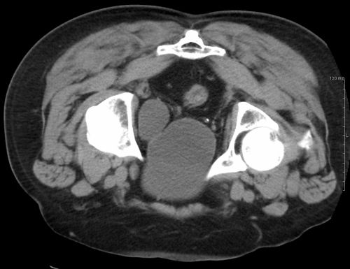 Axial CT scan demonstrating a bladder diverticulum. Source: Wikipedia
- Müllerian cysts
- Uterine, ovarian, and fallopian tube abnormalities
- Urachal cysts
- Ectopic ureter or ureterocele
- Postsurgical changes, including lymphocele
- Lower urinary tract imaging
- Can include cross-sectional studies and/or VCUG
- Cross-sectional imaging may provide useful information regarding bladder diverticula (number, location, filling-defect in case of obstructed diverticulum neck) and critical information regarding the surrounding anatomy.
- VCUG with anterior-posterior, oblique, and lateral images provides information regarding anatomy, location, and size and also associated vesicoureteral reflux and, importantly, emptying of the bladder diverticulum with voiding
- Can include cross-sectional studies and/or VCUG
- Consider upper tract imaging
- Up to 7% of adults with bladder diverticula have been found to have asymptomatic or silent hydroureteronephrosis related to the diverticulum
- Up to 30% of children with bladder diverticula have been found to have upper tract abnormalities, including renal scarring, dysplasia, and hydronephrosis

Other[edit | edit source]
Cystoscopy[edit | edit source]
- The entire interior of each bladder diverticulum should be thoroughly inspected for stones or abnormal-appearing epithelium.
- Any abnormal-appearing epithelium or lesions within the diverticulum are carefully biopsied
- Biopsy should be performed carefully to prevent perforation because the wall of the diverticulum is very thin owing to the lack of a muscularis propria layer
- Any abnormal-appearing epithelium or lesions within the diverticulum are carefully biopsied
Urodynamics[edit | edit source]
- VUDS can be very helpful in patients with bladder diverticula to potentially identify neurogenic voiding dysfunction.
- Failure to identify and treat an existing underlying urodynamic abnormality prior to or concomitantly with definitive surgical therapy for bladder diverticula may result in a high risk of recurrent diverticula or other problems following surgery.
- Bladder contractility may appear to be diminished on urodynamics because of the “pressure sink” effect of the bladder diverticulum.
- This artifact occurs as the detrusor contracts and the intravesical contents are decompressed through the path of least resistance into the bladder diverticulum as opposed to the urethra. Nevertheless, when carefully measured, bladder contractility is not significantly different from that in individuals with benign prostatic hyperplasia without bladder diverticula. In addition, elevated postvoid residual urine may be present as a result of retained urine in the diverticulum regardless of the presence or absence of impaired bladder contractility or bladder outlet obstruction.
Management[edit | edit source]
Options[edit | edit source]
- Surveillance
- Surgery (endoscopic, open, laparoscopic, robotic)
- Catheterization
Surveillance[edit | edit source]
- Suitable for adult patients with minimal symptoms and no complicating factors
- Patients should be counseled regarding the potentially increased risk of malignancy and the need for periodic reassessment as well as the unpredictable course and potentially aggressive nature of malignancy if subsequently found in this setting.
- The association between bladder diverticula and bladder cancer has been attributed to urinary stasis and to chronic inflammation that is often found on pathologic examination of bladder diverticulum tissue.
- Historically, prophylactic surgical treatment was done for all bladder diverticula, including asymptomatic and minimally symptomatic lesions. However, more recently it has been suggested that such management is not warranted, and observation with careful follow-up and periodic endoscopic surveillance is acceptable in asymptomatic individuals without evidence of dysplastic or malignant changes
- The optimal schedule and type of surveillance for those individuals choosing observation is not well defined but consists of:
- Periodic reassessment of symptoms
- Urine studies (including cytology)
- Endoscopic examination of the lower urinary tract
Surgery[edit | edit source]
- Indications for intervention of bladder diverticulae (5):
- Stones in diverticulum
- Upper urinary tract deterioration as a result of obstruction or reflux
- Persistent symptoms not otherwise responsive to medical therapy
- Recurrent UTI
- Carcinoma or premalignant change
- Neoplasms within a bladder diverticulum are associated with poor prognosis because of the potential for rapid transmural involvement of invasive bladder cancer and extravesical extension due to the bladder diverticulum wall lacking a well-developed muscularis propria layer
- The lack of a defined muscular wall risks early dissemination of tumor cells into an extravesical location during transurethral resection of these lesions and makes precise pathologic staging difficult. Because pathologic staging following transurethral resection is difficult and often inaccurate, consideration should be given to the possibility of early extravesical disease extension owing to the lack of a defined muscularis propria and an aggressive approach to these tumors has been suggested.
- ***2015 CUA NMIBC Guidelines include “invasive tumours involving bladder diverticula” as indication for timely cystectomy
- Size of the diverticulum does not correlate with symptoms or complications (UTI or malignancy) and therefore cannot be used as an absolute indication to proceed with surgery
- In patients with bladder diverticulum secondary to bladder outlet obstruction, management of bladder outlet obstruction should occur prior to or concomitant with treatment of the bladder diverticulum. Whether such procedures should be done concomitantly or staged is controversial
Endoscopic surgical management[edit | edit source]
- Transurethral resection of the diverticular neck may be combined with fulguration of the entire urothelial lining of the diverticulum
- The neck of the diverticulum is incised using the resectoscope loop or Collins knife. Incisions are carried down to the muscular fibers of the bladder at the level of the ostium of the diverticulum. Circumferential resection of the entire neck may be performed.
- Fulguration of the lining of the diverticulum should result in obliteration of the diverticulum or a considerable reduction in its size.
- May be considered in:
- Elderly or comorbid
- Those who are not good candidates for open operative approach
- Those undergoing TURP in whom there is an associated poorly draining diverticulectomy
Open/laparoscopic/robotic surgical management[edit | edit source]
- Careful dissection is required during excision of the diverticulum to avoid ureteral injury, because many bladder diverticula are located adjacent to the ureter or may be adherent to it. Ureteral stents are often placed preoperatively or intraoperatively to facilitate dissection and avoid ureteric injury
- Open
- Usually performed through a transvesical, extra-peritoneal approach
- Adverse Events
- Intra-operative:
- Ureteral injury
- Bleeding
- Injury to adjacent organ
- Early post-operative
- Bleeding
- UTI
- Prolonged urinary extravasation postoperatively
- Late-post-operative:
- Urinary fistula
- Intra-operative:
Catheterization[edit | edit source]
- Patients who have poor bladder emptying following relief of obstruction and remain symptomatic, or those who are unable or unwilling to undergo surgical excision of the bladder diverticulum, may be effectively treated with CIC or an indwelling catheter.
Questions[edit | edit source]
- Where are most congenital bladder diverticuli located?
- What is the work-up of an adult patient with a bladder diverticulum?
- List causes of bladder diverticulae in women
- List differences between congenital and acquired diverticulae
- What are the indications for intervention of a bladder diverticulum?
- What are potential complications related to repair of bladder diverticula?
Answers[edit | edit source]
- Where are most congenital bladder diverticuli located?
- Ureterovesical junction
- What is the work-up of an adult patient with a bladder diverticulum?
- History and physical (DRE if male)
- Laboratory: urinalysis, cytology, PSA (if appropriate)
- Imaging:
- Upper tract: US to rule out hydro
- Lower tract: cross-sectional (CT) +/- VCUG
- Other
- Cystoscopy
- UDS: rule out neurogenic voiding dysfunction
- List causes of bladder diverticulae in women
- Dysfunctional voiding
- Urethral stricture
- Vaginal prolapse
- Bladder neck hypertrophy
- Iatrogenic obstruction resulting from anti-incontinence surgery
- List differences between congenital and acquired diverticulae
- Congenital: no risk of cancer, smooth-walled bladder, may not be associated with obstruction
- Acquired: risk of cancer, thick-walled, trabeculated bladder, almost always associated with obstruction
- What are the indications for intervention of a bladder diverticulum?
- Persistent symptoms not responsive to medical therapy
- Recurrent UTI
- Stones within a diverticulum
- Suspicious of malignancy
- Upper tract deterioration from ipsilateral ureteric obstruction or reflux
- What are potential complications related to repair of bladder diverticula?
References[edit | edit source]
- Wein AJ, Kavoussi LR, Partin AW, Peters CA (eds): CAMPBELL-WALSH UROLOGY, ed 11. Philadelphia, Elsevier, 2015, chap 90